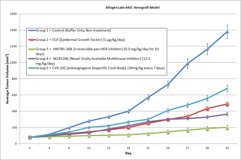
A431 Cell Line Derived Xenograft
A431 is a human epidermoid carcinoma cell line that was originally derived from a skin biopsy of a patient with vulvar squamous cell carcinoma. It is commonly used as a model for studying the biology of epithelial cancers, particularly those of the skin and mucous membranes.
The A431 cell line has several characteristics that make it a useful tool for cancer research. It is able to grow in culture and exhibits many of the same properties as cancer cells found in patients. Researchers have used the A431 cell line to study the molecular mechanisms of epithelial cancers, including the genes and pathways involved in cancer cell growth, invasion, and metastasis.
Angiogenesis is a requirement for tumor take and growth in a xenograft mouse model. The A431 xenograft (CDX) exhibits high secretion of VEGF and is therefore a key model used in novel mono-therapies (e.g. phenylacetate carboxymethyl benzylamide dextran), combination therapies (e.g. gefitinib and cetuximab) or in the direct targeting of EGFR (e.g. 14E1).
| A431 | p53(mut) |
| Origin | Skin |
| Disease | Carcinoma, epidermoid |
| Metastatic Models (Melanoma) | A2058, A375, B16 |
| Non-Metastatic Models (Melanoma) | A431, SK-MEL-2 |
Get Instant Quote for
A431 Xenograft Model
What is a Xenograft?
Development of an anti-cancer therapeutic requires intense, well planned studies that follow a streamlined path for success. Primary studies are performed in an in vitro setting that allows for high throughput screening and analysis of multiple compounds of interest. This method enables a focused compound screening approach of multiple cell lines within a specific cancer type, or a divergent approach across a broad range of cancer types. Ultimately, in vitro screening results need to be confirmed in an animal model due to in vitro inadequacies of cells cultured on plastic, as this method is far removed from the microenvironment of a tumor.
As the logical next step in therapeutic development is the administration of the test compound in a living animal, a cell line derived xenograft model (CDX) is created by inoculating human cancer cell lines in test animals. The injected cell lines grow into established tumors, thus, permitting efficacy studies of the test compounds. An alternative to CDX models is the patient derived tumor xenograft (PDX) which consists of implanting human tumor fragments directly in a mouse model. The PDX model avoids concerns with the CDX model since the tumor is never grown on plastic and there is no selection for single cell populations. In contrary to CDX models, the ideology of PDX models is to maintain the cell population, structure and stroma of the initial tumor.
Why use Xenograft Models?
Cell line derived xenograft (CDX) models or patient derived tumor xenograft (PDX) models enable a larger realm of parameters to be studied not capable with in vitro studies. The complete animal system model expands the scope of studies available to include the effect of test compounds on pharmacokinetics (PK), pharmacodynamics (PD), alternate routes of delivery, inhibition of metastasis, CBCs, dosing regimens, dose levels, etc. However, one of the major drawbacks of CDX and PDX models is that the human cancer cell lines or human patient derived tumors must be implanted in immunocompromised mice in order to bypass the graft versus host rejection by the animal. With the increasing focus of the immune systems role in the recognition and elimination of tumor cells (i.e. immunotherapy), major consideration must be taken into account during experimental concept design of the limitation of checkpoint inhibitors or desired immune response involvement in tumor efficacy. Similarly, any tumor regression after treatment with a test compound in these models will not exhibit the potential complement cascade or innate immune response of the injected therapeutic in humans.
What we offer?
Our in vivo xenograft service department evaluates the efficacy of preclinical and clinical cancer therapeutics utilizing more than 50 validated immunocompromised xenograft mouse models. The value of utilizing our xenograft service department is highlighted by the ability to completely characterize the efficacy, dose regimen, dose levels and optimal combination ratios of lead compounds for cancer, obesity, diabetes, infections and immunology research.
During the design and execution of the xenograft study, our scientists will communicate with and assist the client’s decisions regarding these details:
- Study Group Formation: classification of mice by body weight, tumor size or other parameters
- Cancer Cell Line: use of in-house cell lines or utilization of customer-provided cell lines
- Tumor Implantation: intraperitoneal, subcutaneous, submuscular or intravenous
- Test Compound Administration: intraperitoneal, intravenous, tail vein, subcutaneous, topical, oral gavage, osmotic pumps or subcutaneous drug pellets
- Sample Collection: Tumors/tissues can be fixed in 10% NBF, frozen in liquid nitrogen, or stabilized in RNAlater; blood chemistry analysis can be performed throughout the in-life portion of study